





 Monday, 12-10-26








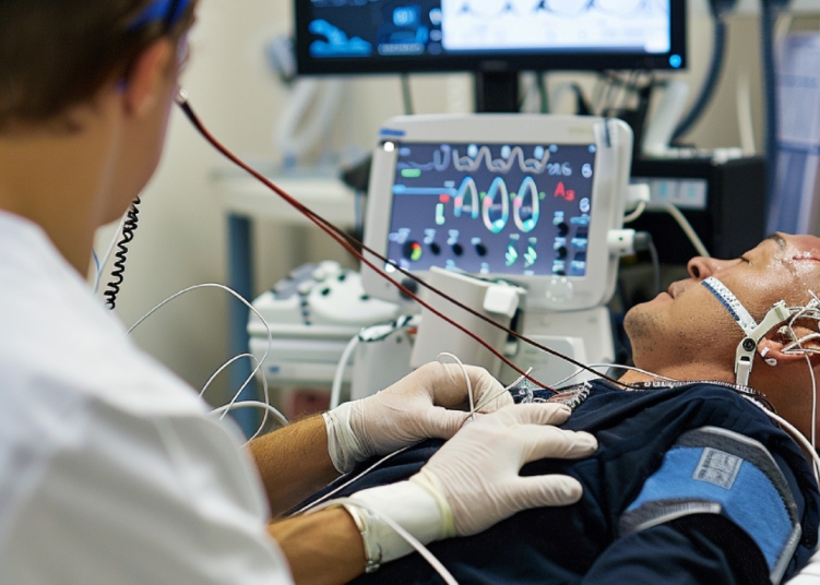
Coronary artery disease remains one of the leading causes of morbidity and mortality worldwide, presenting intricate clinical challenges for interventional cardiologists. As fatty deposits and plaque accumulate within the coronary arteries, blood flow to the heart muscle becomes severely restricted, increasing the risk of angina, heart failure, and life threatening heart attacks. In standard cases, interventional cardiologists rely on traditional percutaneous coronary intervention, commonly known as balloon angioplasty and stenting, to reopen narrowed vessels. However, as coronary artery disease progresses, plaque deposits often undergo extensive calcification, transforming soft arterial buildup into rigid, rock hard calcium structures. Treating these heavily calcified lesions requires specialized advanced techniques, where the Rotablator heart procedure plays a pivotal role in modern cardiovascular care.
When coronary arteries become heavily calcified, conventional balloon catheters and metallic stents encounter significant operational limitations. Inflatable balloons may fail to expand against rigid calcium walls, and attempting to force expansion can lead to severe complications such as arterial dissection, balloon rupture, or incomplete stent deployment. An under expanded stent significantly raises the long term risk of restenosis, where the vessel narrows again, or stent thrombosis, which can trigger sudden cardiac events. To address these complex anatomical obstacles, leading cardiovascular centers utilize the Rotablator heart procedure to safely clear rigid blockages and restore optimal vascular function.
Understanding Complex Coronary Artery Disease And Severe Calcification
Coronary artery disease develops over decades through a gradual process known as atherosclerosis. Initial arterial buildup consists primarily of soft lipids, cholesterol, and inflammatory cellular debris. Over time, chronic inflammation and cellular degeneration cause calcium salts to deposit within these atherosclerotic plaques. As calcification intensifies, the affected coronary blood vessels lose their natural elasticity and become extremely stiff. Severely calcified lesions are particularly prevalent among elderly individuals, patients with long standing diabetes mellitus, and individuals suffering from chronic kidney disease.
Managing calcified coronary blockages requires precise diagnostic evaluation and tailored therapeutic strategies. Standard diagnostic tools like traditional coronary angiography provide two dimensional views of blood flow but may not fully reveal the true depth, volume, or circumferential extent of calcium deposits. When an interventional cardiologist encounters a blockage that cannot be dilated with standard low pressure or high pressure balloons, proceeding with standard stenting carries unacceptable clinical risks. Unmodified calcified tissue acts as a physical barrier, preventing the metallic stent from fully expanding and conforming to the arterial wall. This clinical dilemma underscores the necessity of specialized plaque modification technologies designed specifically for hard tissue debulking.
How Rotational Atherectomy Overcomes Resistant Arterial Plaque
The Rotablator heart procedure, technically referred to as rotational atherectomy, represents a remarkable technological breakthrough in catheter based cardiac interventions. Unlike conventional angioplasty that relies on hydraulic pressure to push plaque outward against vessel walls, rotational atherectomy actively removes and modifies hard calcified tissue from inside the lumen. The system utilizes an ultra thin flexible catheter tipped with an elliptical diamond coated burr. Driven by a compressed air turbine, this microscopic burr rotates at extraordinary speeds ranging from 140,000 to 190,000 revolutions per minute.
As the high speed rotating burr gently advances through the calcified coronary segment, it differential cuts rigid calcium while safely deflecting off soft, elastic arterial tissue. The microscopic diamond grit pulverizes the hard calcified plaque into microparticles measuring less than five microns in diameter, which is smaller than a typical human red blood cell. These tiny particles travel harmlessly through the microvascular capillary bed and are naturally filtered out by the body's reticuloendothelial system without causing distal embolization or tissue ischemia. By shaving down the calcified obstruction, the Rotablator heart procedure transforms a rigid, unyielding vessel segment into a flexible, compliant channel. Once plaque modification is complete, interventional cardiologists can easily glide standard angioplasty balloons and deploy drug eluting stents, ensuring complete vessel expansion and restoring unobstructed blood flow to the myocardium.
The Critical Role Of Intravascular Ultrasound In Precision Cardiology
The success of rotational atherectomy is significantly enhanced when paired with advanced intracoronary imaging technologies, particularly Intravascular Ultrasound, known as IVUS. While traditional angiography provides an external shadow view of the blood vessel, IVUS utilizes a specialized catheter equipped with a miniature ultrasound transducer to generate detailed, three dimensional, cross sectional images from inside the coronary artery. This advanced diagnostic tool enables cardiologists to examine the internal vessel architecture, evaluate the precise thickness and arc of calcium, and measure exact luminal dimensions in real time.
Prior to performing the Rotablator heart procedure, interventional specialists use IVUS guidance to assess whether rotational atherectomy is the most appropriate intervention for the patient's unique vascular anatomy. IVUS imaging helps determine the precise size of the diamond burr required for safe plaque debulking, minimizing the risk of vessel injury. Following plaque modification and stent deployment, IVUS is performed again to verify that the stent has fully expanded, apposed tightly against the arterial wall, and achieved optimal luminal gain. The integration of high resolution imaging with mechanical atherectomy ensures maximum precision, reduces procedural complications, and substantially improves long term patient outcomes in complex interventional cases.
Clinical Benefits And Safety Protocols For High Risk Cardiac Patients
Safety and clinical efficacy are paramount considerations in treating high risk cardiovascular patients with complex coronary anatomy. The primary advantage of rotational atherectomy lies in its unique ability to convert previously untreatable or high risk calcified blockages into routine, manageable stenting procedures. By meticulously modifying rigid plaque, cardiologists avoid applying excessive radial pressure with high pressure balloons, thereby minimizing the likelihood of acute vessel tears, coronary perforations, or severe arterial trauma.
Furthermore, achieving full stent expansion through proper plaque preparation significantly improves drug delivery from drug eluting stents into the vessel wall. Complete expansion reduces the risk of long term complications, such as cardiac reintervention or localized clot formation. Comprehensive preoperative evaluations, including detailed medical history reviews, kidney function assessments, and hemodynamic monitoring, ensure that patients selected for rotational atherectomy receive the safest possible care tailored to their overall health status.
Comprehensive Cardiovascular Centers And Modern Emergency Intervention
Delivering world class care for complex cardiac cases requires advanced medical infrastructure, cutting edge technology, and highly experienced multidisciplinary medical teams. Leading medical institutions, such as Mayapada Hospital through its specialized Cardiovascular Center, have integrated rotational atherectomy and advanced intravascular imaging into their core clinical offerings across major locations including Jakarta, Tangerang, Bogor, Bandung, and Surabaya. These comprehensive centers offer round the clock emergency response services, including specialized 24/7 Cardiac Emergency units operating under international door to balloon protocols aimed at restoring coronary blood flow within critical timeframes.
Equipped with modern cardiac catheterization laboratories, intensive cardiovascular care units, and advanced surgical suites, integrated health systems ensure seamless continuity of care. Patients presenting with intricate, multi vessel coronary disease benefit from collaborative multidisciplinary care teams, comprising interventional cardiologists, cardiothoracic surgeons, cardiac anesthesiologists, and specialized intensive care nurses. By combining state of the art innovations like the Rotablator heart procedure with holistic, patient centered care, modern healthcare providers continue to redefine standards in cardiac treatment, offering renewed hope and improved quality of life for individuals facing complex heart conditions.
Read More